Stena
Stent Intracranial Current Converter
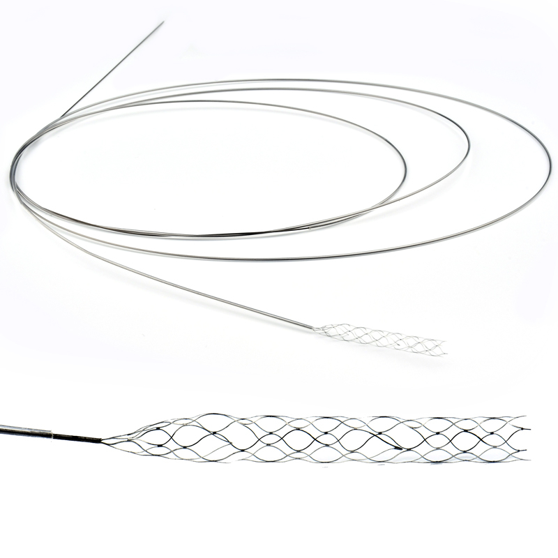
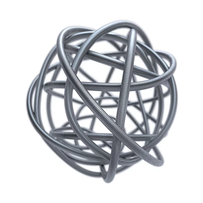
It is a multilayer flow-directed radiopaque tubular self-expandable implant. It consists of multi-layer braided wires made of nitinol. It is designed to be inserted via a percutaneous arterial approach with the StenaⓇ STENT Microcatheter System (Microcatheter 3.6F or 3.9F and Internal MicroCatheter 2.3F). It comes pre-loaded on the delivery system. Microcatheter system supplied separately
The Unique Capabilities That Benefit Patients
Multilayer Flow-guided device is indicated for the endovascular treatment of patients with an intact intracranial aneurysm or pseudoaneurysm (saccular, fusiform, dissection) and morphology suitable for endovascular repair
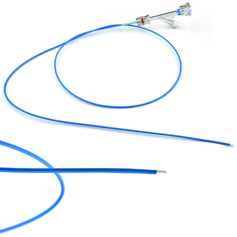
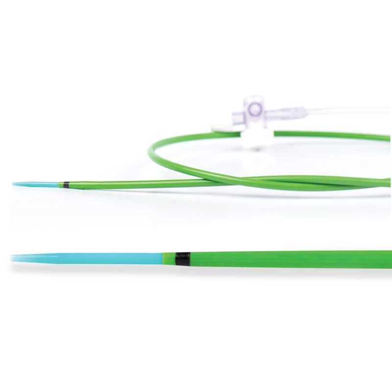
The stent is made of super-alloyed nitinol wire with a self-expandable system, braided in a multi-layer tubular mesh structure. This design structure allows the stent to be flexible, conformable, and self-expanding. There are radiopaque markers made of tantalum at both ends of the stent. It is used with the Microcatheter System. The Microcatheter 3.6F or 3.9F and the Internal Micro Catheter 2.3F are designed to be inserted via a percutaneous arterial approach. It is preloaded in a cartridge in the mesh stent delivery system. The microcatheter system is supplied separately.

Detailed specifications
The StenaⓇ Stent, a Multilayer Flow-guided device is indicated for the endovascular treatment of patients with an intact intracranial aneurysm or pseudoaneurysm (saccular, fusiform, dissection) and morphology suitable for endovascular repair. It can be used as a single device to reconstruct the common artery.
The conduction system consists of a hydrophilic reinforced conduction wire with a radiopaque atraumatic distal end. It is positioned between 2 markers, distal and proximal.
It can treat unruptured intracranial aneurysms or pseudoaneurysms such as saccular, fusiform, and dissection.
Thanks to its multi-layered braided structure, it transforms turbulence into lamination.
The meshwork of the stent influences the hemodynamics of flow in the aneurysm.
As the density of the stent in the mesh increases, the eddy current in the aneurysm decreases.
It accelerates endothelialization with its high density and low pore diameter.
STABILIZATION OF ANEURYSM
- REGULATED THROMBUS
- SIDE BRANCH SPACE
- ENDOTELIZATION
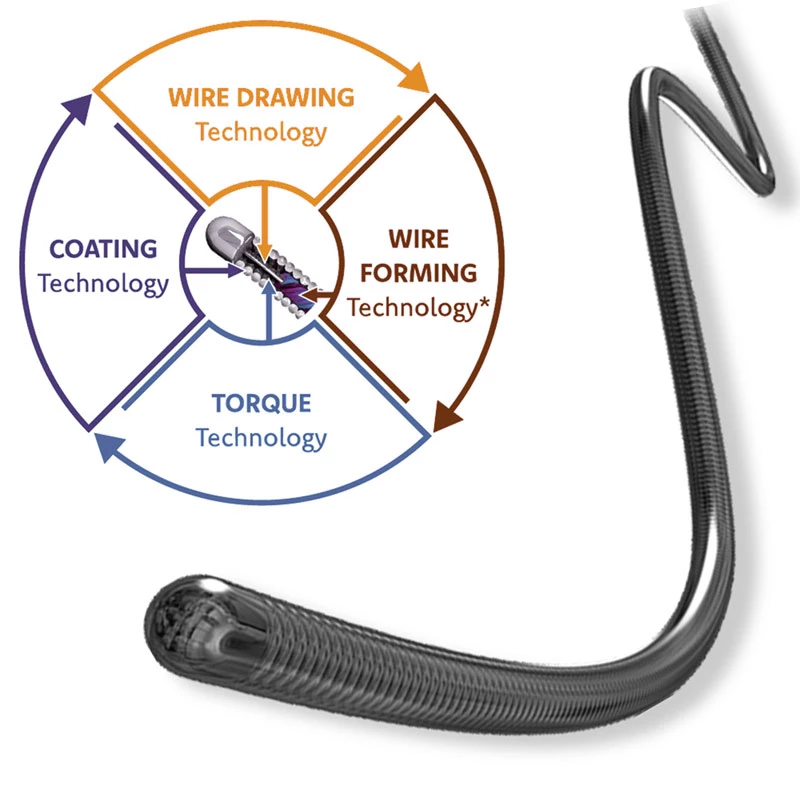
It has a unique structure and flexibility that allows optimum navigation with its nitinol structure:
- High flexibility and conformity
- Visible on MRI
- Hemo compatible surface
- Visible on X-ray
- 3D interlocking braided design
DELIVERY SYSTEM FEATURES
- Easy Distribution
- Hydrophilic distribution wire
- Recapture system
- Atraumatic radiopaque distal helix
MICROCATHETER SYSTEM FEATURES
- Special hydrophilic microcatheter
- Internal hydrophilic micro-catheter provided
- Aneurysm enlargement is the result of tension and loss of tensile strength (TS) in the arterial wall. Rupture occurs when the shear stress exerted by the flow is greater than the resistance of the thinned wall.
- High flexibility and conformity
- Visible on MRI
- Hemo compatible surface
- Visible on X-ray
- 3D interlocking braided design
|
Stent Diameter |
3.0 mm – 5.5 mm |
|
Stent Lenght |
20 mm –80 mm |
|
Catheter Diameter |
6F |
|
Catheter Lenght |
160 cm |
|
Radiopasity |
Pt-Ir Marker |
|
Applicable Microcatheter |
0.027” ID |
|
Stent Design |
Niti Mesh Design |